


In existence since 1975, but open to the public since less than 5 years ago, the Brain Museum in the southern Indian city of Bangalore, encourages visitors to touch its exhibits and ask as many questions about the inner workings of the brain. 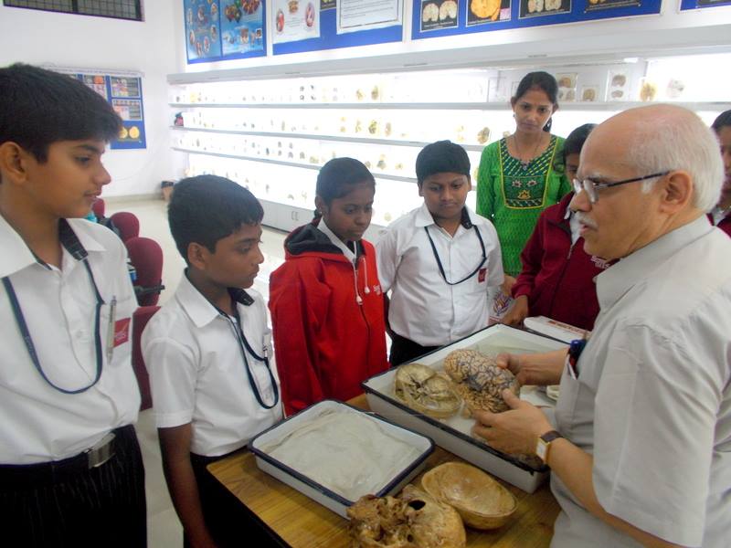
Seen here in the photo, Dr. Prof. S.K. Shankar, who has been a professor at the Department for the past 32 years. Photo courtesy NIMHANS.
The Human Brain Museum is housed at the Neurobiology Research Centre of the National Institute of Mental Health and Neuro Sciences (NIMHANS) in Bangalore, India.
Rereeti spoke to the present co-ordinator at the museum, Dr. S.K. Shankar to dig deep into the workings of the Brain Museum’s history, exhibits, current focus, and its take on revitalizing a techno-medical space.
Rereeti: When and How Was the Brain Museum Set Up?
Museum: Since 1975, the Department of Neuropathology at the National Institute of Mental Health and Neuro Sciences (NIMHANS), Bangalore, has been archiving and mounting brain specimens in various stages of diseases. These mounted specimens are being utilized as a resource for training students of Neurosurgery and Neurology (post-doctoral course). The visitors, usually scientists, doctors, and faculty members from other institutions, used to visit this specimen lab for teaching purposes and also as a resource for examinations. Subsequently, since 2010, this archival collection was formally made accessible to the visitors.
Rereeti: Who is the Primary Audience at the Human Brain Museum?
Museum: The primary audience are students of Neurology and Neurosurgery, Pathology, Psychiatry, Neurosciences, and college and school students. The mission of the Human Brain Museum is to sow the seeds of Neuroscience in school and college students so that they have the general understanding, awareness and choice of career.
Rereeti: Visitors Can Actually Touch the Exhibits at the Brain Museum. This is Rather Unique When it Comes to Museums, Right?
Museum: As far as we know, anatomical museums in general don’t really allow visitors to touch the exhibits. Anatomy specimens are preserved and kept behind glass cases, in cupboards or storage. That’s why the public always has a two dimensional experience of engaging with such exhibits. They usually see pictures or watch videos of the brain and the human body parts, never really gaining an in-depth understanding of the surface, texture, weight, or finer details.
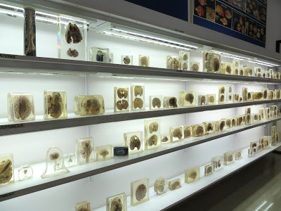
Keeping this in mind, specimens at our museum are mounted in ingeniously made plastic jars designed at our workshop. This has facilitated the viewers to hold the specimens in hand and rotate them in all directions and get an idea of the structure of the anatomy. In addition, depending upon the availability of museum personnel, we provide in-depth explanations to school and college students about the specimen they are interested in. In fact, many of the museum’s foreign visitors expressed that their knowledge of human anatomy and the brain in particular, is based on books and electronic media; they have never had access to real specimens.
Rereeti: What are Some of the Interesting Exhibits Featured at the Museum?
Museum: The museum has sections related to head injury, cerebrovascular diseases, common neuro-infections involving the brain, developmental disorders of the brain, and brain tumors. The cerebral vessels supplying the brain are dissected in normal and pathological states and mounted to visualize the normal anatomy and its alterations in pathological states. In addition, many posters related to Epilepsy, Autism, organ donation, general information about the nervous system are displayed and the public are permitted to take photographs. During guided tours, visitors are made aware of treatable conditions, curable conditions and the diseases that cannot be cured.
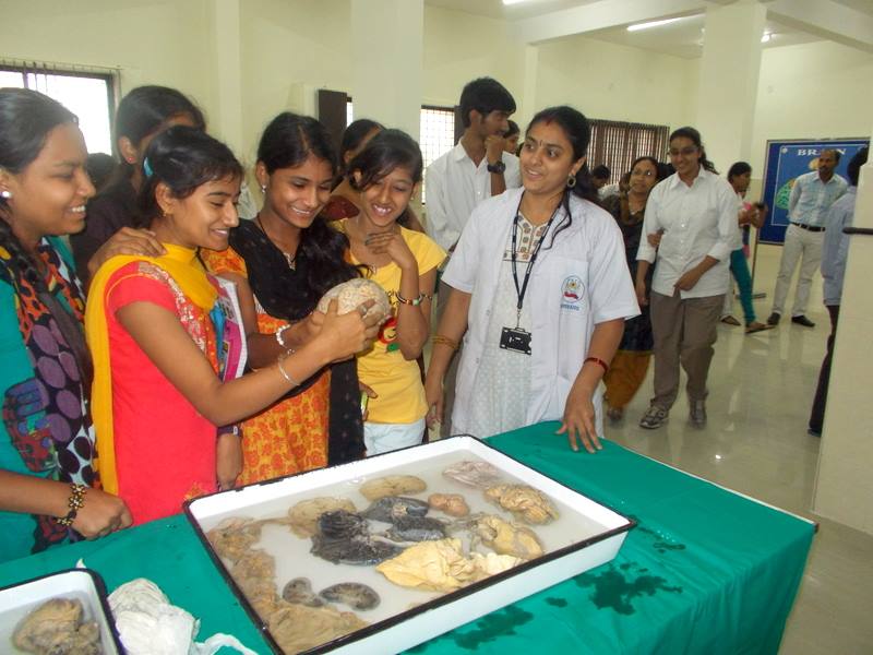
Image: College students handling a brain specimen at the Museum. Photo courtesy: NIMHANS.
Rereeti: How Do You Make Science, Particularly Anatomy, Accessible to the Public in Terms of Content, Presentation and Relevance?
Museum: The first part about accessibility comes with touch. At our museum, visitors have first-hand experience of engaging with the exhibits as they can touch and feel the human brain, heart, lung, kidney, spinal cord, intestine, pituitary gland, and other organs that are displayed in their original form (formalin fixed, non-contagious and safe to handle). In terms of content relevance, since we organize guided tours for school and college students, they get in-depth knowledge related to their curriculum. We also focus on altered anatomy in diseased states, especially life style related disorders. In addition, a few charts are available.
Rereeti: What Are Some of the Challenges Facing the Museum?
Museum: The foremost challenge faced by the Human Brain Museum is the non-availability of permanent staff or trained volunteers to organize guided tours. We maintain ourselves through the generous, one-time support of the NIMHANS administration. However, lack of funds for upkeep, maintenance and museum personnel is making it difficult to revitalize the museum, plan alterations or improvements. Space is also a vital constraint.
Rereeti: What is Your Wishlist for the Museum?
Museum: Presently, with the limited funding and minimal staff, we are hesitant to dream. Unlike other large museums, the Human Brain Museum is unique and needs personal interactions between the co-ordinators and the public to make the museum visit meaningful. If a larger space is provided and with commensurate funding, there are additional brain specimens to be mounted, along with the idea of curating animal brain specimens for comparative Neuro-anatomy and for research on evolution of nervous system.
Possibly this knowledge can provide insight into understanding of nervous system and take it to a translational research, applicable especially to the field of neurodegeneration and stem cell research for many of the presently untreatable conditions. This will facilitate not only making the brain museum a space of tourist attraction but a place of scientific resource. This is doable with the commitment of space, funds and attracting a few committed scientists.
Also on the wishlist are audio-guided tours and interactive touchscreen kiosks to help reduce dependency on volunteers from time to time. We would also love to digitize our museum collection. A limited number of permanent staff with respectable salary are needed to pursue this as a career opportunity and a long-term commitment.
About the Author
Dr. S.K.Shankar MD  Emeritus Professor, Neuropathology (retired as Prof & Head –Dept. of Neuropathology in 2012). Principal Co-Ordinator- Human Brain Museum & Brain Bank, NIMHANS, Bangalore. Evolved the Human Brain Museum with the participation of faculties of department of Neuropathology for the past 30 years and transformed to present Neuropathology Museum at Neurobiology Research Centre, NIMHANS-Bangalore. Managing the Human Brain Museum and actively participating in teaching the medical students and general public with the assistance of other member faculty of Department of Neuropathology.
The museum is open for guided tours on Saturday between 9 a.m. and 4.30 p.m. and on Wednesdays between 2 p.m. and 5 p.m.
Reblogged this on EVE Museografía.